|
|
Korenine / Origin / Herkunft |
|


| © korenine.si | Podpora |
|
||||||||||||||
| Origin | Conference | News | Proceedings | About us | Links | Donations |
|
|
|
3D FACIAL RECONSTRUCTION FROM A SKULL OF A MALE SUBJECT OF THE NEOLITIC SQUARE MOUTH POTTERY CULTURE OF QUINZANO (VERONA, ITALY)
M. Silvestri1, Giancarlo Tomezzoli2
Abstract In this paper we used a 2D - 3D computer graphics technique to reconstruct a 3D model of the face of a Neolithic male subject belonging to the Square Mouth PotteryCulture. The skeleton and the skull of the subject were found near Quinzano (Verona, Italy) and are preserved at the Civico Museo di Storia Naturale in Verona (Veneto, Italy). Despite the skull’s bad condition, we were successful in restoring its main anthropometric facial features by designing a three-dimensional skull model, and by creating a final ray-tracing image representing the face as it may have looked when the subject was alive. The result, despite the technical limitations of our method, leaves no doubts that the subject was of European origin, and the reconstructed face appears to be very common in northern Italy and central Europe’s present-day male subjects.
Introduction - 3D Facial Reconstruction Methods.A traditional technique of reconstructing a face from a skull is to start from a pre-made plastic model and then adding the flesh layers one by one, according to the famous technique used by Prag and Neave [1]. With the enormous progress, in the recent decades, in the fields of the Computer Graphics it is now possible to recreate the human face from an ancient skull in several ways, i.e.: from a sequence of photographic skull image frames [2,3,4], from a 3D skull model obtained by using a Computed Tomography (CT) scanning technique [5], from a 3D skull model based on voxels [4], simply by modelling warping points on the skull and adding the soft tissue features [6, 7]. Whatever the technique used, the results are generally good and respect the main skull features. The main problem is related to the calculation of the exact flesh thickness, and how to guess a realistic pigmentation for skin, eyes, and hairs. In our present work we use a hybrid 2D – 3D stereo imaging [3]technique to create a 3D skull model on which we added the soft tissues. For the thickness of the soft tissues we used values form Prag and Neave [1,8] which already proven successful in archaeology and forensic anthropology and were used by many other authors [9]. In order to test the reliability and the possible general applicability of this hybrid facial reconstruction technique, we decide to apply it to the skull of the skeleton of a male subject who lived in the area of the today town of Quinzano (Verona – IT), aged of about 6000 yr. The skull and the skeleton (cf. Figure 1) are preserved at the Civico Museo di Storia Naturale in Verona.
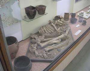
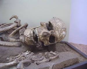
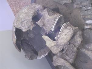
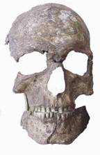
3D Facial Reconstruction – Application & ImplementationIn Figure1 is shown the skeleton and the skull of the male subject of the so-called Neolitic “square mouth vases” Culture. As set out in the boards not far from the skeleton, this culture developed in three distinct phases: Quinzano, Rivoli-Chiozza, Rivoli-Castelnuovo. The settlement of Quinzano was an open air one. The excavations made by Zorzi have revealed an extended village with a necropolis of buried, crouched peoples. This settlement is typical of the first phase of the said “square mouth vases” Culture, which had, as distinctive feature, glasses and vases with a square mouth, decorated with simple geometrical motifs. Some square mouth artefacts are visible in Figure 1 near the skeleton. The front side and the lateral side of the skull are shown in Figures 2 and 3.
Figure 1. Skeleton from the “square mouth vases” Figure 2. Skull – front view. culture, Museo di Storia Naturale, Verona (Italy).
Figure 3. – Skull – side view. Figure 4. – Jaw and nose bridge corrections
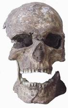
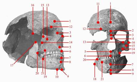
The set of photos in Figures 1-3 was produced by means of a digital camera and provide a set of high-resolution photographic images suitable for facial reconstruction. At first glance it appears that the skull is largely incomplete, but, fortunately, not in parts concerning directly the face. The subject was a male, probably 25-30 years old. The causes of the death, apparently, cannot be determined. As shown in Figure 4, the skull was isolated from its context, some not essential features were eliminated, and the open jaw was repositioned in its natural closed position by using specific 2D editing software. The same software has corrected the following deformations of the original skull: - incomplete nose bridge; - distorted nose shape, sharply tending to the right side of the skull; - jaw’s missing pieces; - wide open left side skull. The digital images have been processed by using a 2D stereo imaging technique derived from the works of Chen, Medioni, Zhan [2,3] and D’Apuzzo [4], in order to create a correct 3D skull model by using commercial 3D CAD/CAM software. Because the skull of this ancient male subject does not appear very different from the skulls of the present people, and because, as far as can be understood from the images, the subject was not emaciated or obese, it seems reasonable to assume that the thickness of the soft tissues on the different portions of the skull was not dissimilar from that of the today’s Central European people. Therefore, once the 3D model of the skull was finally obtained, we established, according to the technique used in the works by Neave, Prag and Wilkinson [1,6,8,9], a set of warping points 1-21 (cf. Figure 5) having corresponding soft tissue thickness values in a Look Up Table (LUT) for a normal Central European male (cf. Table 1).
Figure 5. Skull warping point set.
In addition, from the images we derived front-skull facial dimensions and proportions to be added where necessary, as further details, in the final 3D facial reconstruction. Despite of all our efforts, some slight imperfections in the reconstructed skull model could interfere in the final 3D facial reconstruction, however, it is believed that said imperfections could not significantly alterate the final 3D facial reconstruction.
Table 1. Table of measurements for flesh thickness, after J.S.Rhine and C.E. Moore, Forensic Anthropology : Maxwell, Museum Technical Series (1984) (from Prag and Neave [1]).
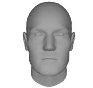
Figure 6 shows the preview of the facial reconstruction derived on the basis of the above-mentioned set of warping points and the corresponding soft tissue thickness listed in the LUT.
Figure 6. Preview of the 3D facial reconstruction.
Because the main goal of our research is to produce a basic and credible 3D facial reconstruction, we avoided the implementation of recent finer techniques as proposed by De Carlo [7].
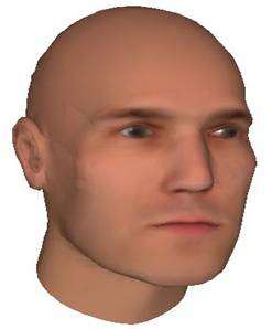
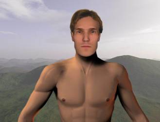
Pigmentation Because no rests of the hair, beard of moustaches or other indicia are present near the skull we cannot have a clear idea of the original pigmentation of the skin, eyes, beard and moustaches if any. However, at least concerning the skin, many statistics on the pigmentation of Europeans are in agreement in considering the Central or Alpine Europeans deprived of a clear dominant pigmentation, and in considering the skin pigmentation varying in the range from very fair to mid dark [10, 11, 12]. Therefore, we proceeded for the 3D final facial reconstruction, by adopting a typical fair skin and brownish eyebrows and eyes as shown in Figure 7. An artistic facial reconstruction of the subject in a landscape similar to that of the actual Quinzano is provided in Figure 8.
Figure 7. Final 3D facial reconstruction Figure 8. Artistic facial reconstruction ConclusionsAs set out above, shape and/or thickness of the soft tissues play a crucialrole in 3D facial reconstruction. However, the absence, at least at the moment, of precise rules for establishing soft tissues shape and/or thickness obliged us, in our opinion, to the reasonable assumption that on the different portions of the ancient skull the thickness of the soft tissues was the same as that of the today’s Central European people. For this reason, the reconstructed face looks not dissimilar from the faces of the today Central European subjects.
Bibliography 1. J Prag, R Neave, Making Faces Using Forensic and Archaeological Evidence, British Museum Press, London 1997 2. Q Chen, G Medioni, Building human face models from two images, IEEE 2nd Workshop Multimedia Signal Processing, Dec. 1998, 117-122 3. Z Zhang, Image-based modelling of objects and human faces, Proc. SPIE, vol. 4309, Jan. 2001 4. N D’Apuzzo, Measurement and Modeling of Human Faces from Multi Images, International Archives of Photogrammetry and Remote Sensing, 2002, 34(5), 241-246 5. M Yoshino, H Matsuda, S Kubota, K Imaizumi, S Miyasaka, S Seta, Computerassisted skull identification system using video superimposition, Forensic Science International, 1997, 90, 231-244 6. P Vanezis, R W Blowes, A D Linney, A C Tan, R Richards, R Neave, Application of 3-D computer graphics for facial reconstruction and comparison with sculpting techniques, Forensic Science International 1989, 42, 69-84 7. D DeCarlo, D Metaxas, M Stone, An Anthropometric Face Model using Variational Techniques, Proceedings SIGGRAPH ’98, 67-74 8. R A H Neave, A J N W Prag, The skull as the armature of the face: reconstructing ancient faces, In: A Bowman, and M Brady (eds.), Artefacts and Images of the Ancient World, British Academy, London 2000 9. C Wilkinson, R Neave, The reconstruction of a face showing a healed wound, J. Archaeol. Sci. 2003, 30(10), 1343-1348 10. L Farkas, Anthropometric Facial Proportions in Medicine, Thomas Books, Springfield 1987 11. L Farkas, Anthropometry of the Head and Face. 2nd ed., Raven Press, New York 1994 12. C S Coon, Races of Europe, Macmillan, New York, 1939
Additional Material The original high-resolution images used for the 3D facial reconstructions and all the produced material is available by the authors: M. Silvestri (marco@marcosilvestri.com), G. Tomezzoli (gtomezzoli@epo.org).
Povzetek 3D rekonstrukcija obraza lobanje moškega pripadnika neolitske kulture posod s kvadratnim ustjem iz Quinzana (Verona, IT) Za rekonstrukcijo 3D modela obraza neolitskega moškega, pripadnika kulture posod s kvadratnim ustjem, sva uporabila 2D - 3D računalniško grafiko. Njegovo okostje in lobanjo so našli pri kraju Quinzano (Verona – IT) in ga hranijo v Civico Museo di Storia Naturale v Veroni (Veneto – IT). Kljub temu, da je v slabem stanju, sva uspela vzpostaviti glavne antropometrične značilnosti obraza tako, da sva pripravila 3D model lobanje in končno s svetlobnim risanjem podobo, ki kaže, kako je morebiti bil videti njegov obraz, ko je bil še živ. Kljub tehničnim omejitvam najinega pristopa je videti rekonstruirani obraz podoben obrazom sedanjih moških v severni Italiji in sploh srednji Evropi.
|